Choledochal Cysts Type I
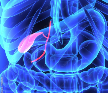
A type 1 choledochal cyst is the most common form of a rare condition, and the one surgeons know best. This page covers what it is, how it is found, how it is treated, and what follows.
What a type 1 choledochal cyst is
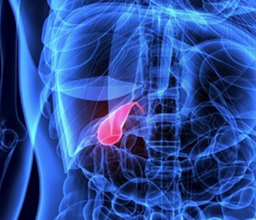
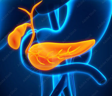
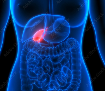
Bile is made in the liver and travels down small tubes — bile ducts — into the intestine, where it helps digest fat. Picture a river system: little streams inside the liver join into two branches, which merge into one main channel outside the liver, the common bile duct. The gallbladder sits on a short side branch.
A type 1 cyst is a balloon-like widening of that main channel outside the liver. It is not a tumour; the duct is simply built too wide from birth, so bile pools instead of flowing through. The streams inside the liver are normal. That detail — swelling outside the liver only — separates type 1 from the other types of choledochal cysts.
Subtypes: type Ia, Ib and Ic
Radiologists split type 1 into three patterns:
- Type Ia — the whole extrahepatic bile duct balloons into a rounded, cystic sac. The classic picture.
- Type Ib — only a focal segment is widened, with normal duct above and below.
- Type Ic — the duct is widened along its length in a smooth, tapering, cigar shape: fusiform rather than cystic.
Types Ia and Ic usually come with a pancreaticobiliary maljunction: the bile and pancreatic ducts meet unusually early, outside the muscular valve that normally keeps them apart. Pancreatic juice can then wash back and irritate the bile duct lining, which is thought to be part of why the duct becomes damaged over time.
How common is it, and who gets it
Type 1 is by far the most frequent shape: published series put it at roughly 50% to 80% of all choledochal cysts, and some radiology sources at 80% to 90%. Types 1 and 4 together make up about 90% of cases.
Choledochal cysts overall are rare in Western countries — roughly 1 in 100,000 to 1 in 150,000 people — but far more common in East Asia, approaching 1 in 1,000 live births. Girls and women are affected about three to four times as often as boys and men. Around 80% of patients come to attention before age 10; only about 25% are first found in adulthood.
How it usually shows up
The textbook classic triad is belly pain, jaundice (yellowing of the skin and eyes), and a lump under the right ribs. All three appear together in only about 20% to 30% of patients; most people have one or two. Babies more often show jaundice and pale stools; older children and adults tend to have recurrent right-sided pain, nausea or pancreatitis. See symptoms.
How it is diagnosed
Ultrasound is almost always first — painless, no radiation, and it picks up a dilated duct in 71% to 97% of cases. It cannot reliably show how the bile and pancreatic ducts join, so the test that settles the diagnosis is usually MRCP, a special MRI of the bile ducts, with reported accuracy of about 90% to 100% and no instruments or radiation. ERCP gives the sharpest pictures but is invasive, so it is usually saved for when treatment can happen at the same time. See diagnosis.
How type 1 cysts are treated
The standard operation is complete excision of the dilated duct plus removal of the gallbladder, with reconstruction by Roux-en-Y hepaticojejunostomy — a loop of small intestine joined directly to healthy duct near the liver. Many centres now do this laparoscopically or robotically.
Why the whole duct comes out
Why remove the duct rather than just drain it? Because the abnormal lining is the problem. Older drainage operations left it in place, and those patients developed bile duct cancer at roughly four times the rate of those fully excised. This is the key difference from a type 3 choledochal cyst, which sits in the duodenal wall and can often be handled endoscopically, and from a type 2 choledochal cyst, where only a small pouch needs removing. Types IV and V involve ducts inside the liver, making surgery more complex. See preparing for surgery and postoperative care.
Complications and cancer risk
Left alone, a type 1 cyst can cause repeated bile infections (cholangitis), stones, pancreatitis, liver scarring and rarely rupture. The concern driving early surgery is cancer. Lifetime malignancy risk across all choledochal cysts is commonly quoted in a range from about 5% up to 30% depending on the series, and type 1 accounts for about 68% of the cancers that arise in them. Risk climbs steeply with age, from well under 1% in the first decade of life to well over 10% by adulthood. See complications.
What follow-up looks like
Excision lowers the risk but does not erase it, because a short stump of duct always remains. Later cancer has been reported in a small minority of patients after excision — around 1.9% after complete excision versus roughly 18% after drainage-only surgery in one large meta-analysis — sometimes more than 15 years later. Lifelong follow-up with liver blood tests and abdominal ultrasound is recommended, plus prompt review for new pain, fever or jaundice. Five-year survival after excision is about 95.5%.
What this means for you
Type 1 is the most treatable version of this condition. The operation is well established, and most children and adults go on to eat and grow normally and live without restriction. Two things matter most: have the cyst fully removed rather than drained, and keep your check-ups for life. To meet families who have been through it, visit find community, or use find doctors.
Note: this page is general information, not medical advice. Please discuss your own situation with your medical team.
Sources
- Choledochal Cyst — StatPearls, NCBI Bookshelf
- Biliary cysts: etiology, diagnosis and management — World Journal of Gastroenterology
- Revisiting the Todani Classification — Diagnostics
- Choledochal cyst — Radiopaedia
- Choledochal Cysts Treatment & Management — Medscape
- Meta-analysis of risk of developing malignancy in congenital choledochal malformation — British Journal of Surgery