Choledochal Cysts Type II
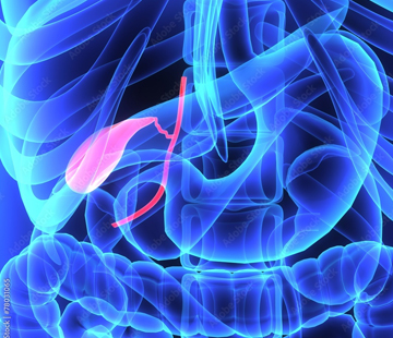
A type 2 choledochal cyst is the rarest of the recognised shapes and, in many ways, the most straightforward. It is a pouch budding off the side of the bile duct rather than a swelling of the duct itself, and that difference changes how it is treated.
What a type 2 choledochal cyst is
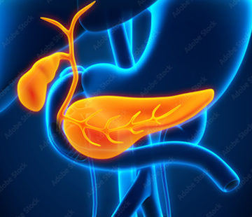
Bile made in the liver drains through small tubes into the intestine. Outside the liver they join into one main channel, the common bile duct, with the gallbladder on a short stalk off its side.
In type 2 the main channel is normal width. Instead a single pouch — a true diverticulum — sticks out from one side, usually above the duodenum, joined by a narrow neck. Think of a small blister on a garden hose: the hose still carries water fine, but a pocket has formed where fluid sits still. Bile stagnates there, and that is where the trouble starts.
Unlike a type 1 choledochal cyst, where the whole duct is dilated, the duct wall in type 2 is healthy, which is why surgeons can take the pouch and leave the plumbing intact. Compare all five shapes on our different types page. Type 2 has no recognised subtypes.
How common is it?
Type 2 is genuinely rare: about 2% to 3% of all choledochal cysts, with some large series reporting 1% to 2%. For context, types 1 and 4 together account for roughly 90% of cases. Choledochal cysts of any kind affect around 1 in 100,000 to 1 in 150,000 people in Western countries, and closer to 1 in 1,000 live births in parts of East Asia, so type 2 is a rare variant of an already rare condition. Many surgeons see only a handful in a career, which is why a specialist centre matters.
Who tends to get it
With so few cases, type 2 has no epidemiology of its own; it is described using the figures for choledochal cysts as a whole:
- Sex — girls and women are affected roughly three to four times as often as boys and men.
- Age — about 80% present before age 10; only around 25% of cases are first found in adulthood.
- Geography — markedly more common in East Asian populations than in Europe or North America.
Unlike types Ia, Ic and IVa, type 2 is not typically linked to a pancreaticobiliary maljunction, the abnormal early meeting of the bile and pancreatic ducts thought to drive damage in the commoner forms.
How it usually presents
Many type 2 cysts are found by accident on a scan done for something else. When symptoms appear they come from stagnant bile or stones forming inside the pouch: right-sided upper abdominal pain, nausea, episodes of jaundice, fever from bile infection (cholangitis), or pancreatitis. The classic triad of pain, jaundice and a palpable mass is reported in only about 20% to 30% of choledochal cyst patients overall, and a small side pouch is far less likely than a large type 1 cyst to be felt as a lump. See symptoms for more.
How it is diagnosed
Ultrasound comes first and detects bile duct abnormalities in 71% to 97% of cases, but it can be hard to tell a type 2 pouch from the gallbladder or a duplication cyst. The decisive test is MRCP, an MRI tuned to show the bile ducts, with reported accuracy of about 90% to 100%. It shows the key finding: a cystic structure communicating with the bile duct through a narrow neck and clearly separate from the gallbladder. ERCP is the most accurate but invasive, so it is reserved for when treatment might happen at the same sitting. See diagnosis.
How type 2 cysts are treated
Treatment is usually simple excision of the diverticulum — a diverticulectomy — with the neck closed off where it met the duct. The defect left in the common bile duct is closed directly, sometimes over a T-tube while it heals.
Why type 2 needs less surgery
In type 1 and type 4 cysts the diseased lining is the duct itself, so the duct must be removed and the system rebuilt with a Roux-en-Y hepaticojejunostomy. In type 2 the duct is healthy and can usually be preserved. A type 3 choledochocele is different again and often managed endoscopically, while type 5 involves the liver itself. Reconstruction is needed in type 2 only if removing the pouch narrows the duct. See preparing for surgery and postoperative care.
Complications and cancer risk
Before treatment the risks are stones, recurrent cholangitis and pancreatitis. Cancer risk is much lower than in type 1: malignant change in types 2, 3 and 5 is described as exceedingly rare, and type 2 accounts for only about 5% of cancers reported in choledochal cysts, against 68% for type 1. The risk is not zero, which is why the pouch is removed rather than watched. See complications.
What follow-up looks like
After a clean diverticulectomy, follow-up is lighter than after a full duct resection but still long-term: periodic liver blood tests and abdominal ultrasound, and a low threshold for imaging if pain, fever or jaundice returns. Reported five-year survival after choledochal cyst excision is about 95.5%.
What this means for you
The honest summary for type 2 is: rare, but usually the simplest to fix. One well-planned operation removes the problem, your own bile duct is normally kept, and the long-term outlook is good. The practical step is finding a surgeon who has handled biliary cysts before — see find doctors — and meeting others who understand the diagnosis at find community.
Note: this page is general information, not medical advice. Please discuss your own situation with your medical team.
Sources
- Choledochal Cyst — StatPearls, NCBI Bookshelf
- Biliary cysts: etiology, diagnosis and management — World Journal of Gastroenterology
- Revisiting the Todani Classification — Diagnostics
- Choledochal Cysts Treatment & Management — Medscape
- Choledochal cyst — Radiopaedia
- Choledochal Cyst — Cleveland Clinic