Choledochal Cysts Type III
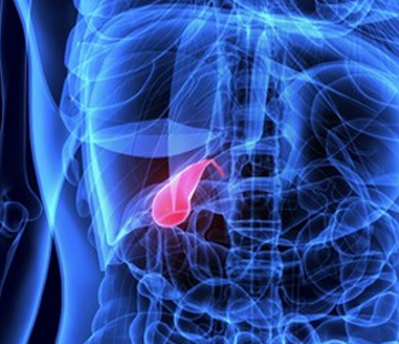
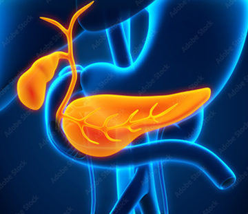
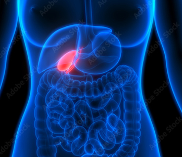
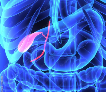
A choledochocele, or type 3 choledochal cyst, is the odd one out. It sits in a different place from the other types, carries a much lower cancer risk, and is usually treated endoscopically rather than with major surgery.
What a choledochocele is
Bile leaves the liver through the common bile duct into the first part of the small intestine, the duodenum. At the very end, the duct tunnels through the duodenal wall and opens at a small bump called the ampulla of Vater, guarded by a ring of muscle (the sphincter of Oddi).
A choledochocele is a balloon-like swelling of that last, intramural segment — the short stretch inside the duodenal wall. Picture a hosepipe passing through a thick wall, with the final inch ballooning inside the wall. On endoscopy it looks like a smooth, soft bulge at the ampulla. Everything upstream is usually normal width, which sets it apart from the other types of choledochal cyst. There are no subtypes.
Some choledochoceles are lined by bile duct (biliary) cells, others by duodenal cells. This matters: a biliary lining carries a higher risk of malignant change, so it is usually biopsied.
How common is it?
Choledochoceles are rare: roughly 1.4% to 5.6% of all choledochal cysts, with most modern reviews quoting 1.4% to 4.5%. A 40-year systematic review covering 1975 to 2015 found just 325 reported patients worldwide. By comparison, type 1 and type 4 together make up about 90% of cases.
Who tends to get it
Type 3 breaks the usual patterns:
- Sex — the strong female predominance seen across choledochal cysts (three to four women per man) is absent in choledochoceles.
- Age — most other types present in early childhood; choledochoceles turn up more often in older children, adolescents and adults. The 40-year review held far more detailed adult reports (58) than paediatric (13).
- Anatomy — an anomalous pancreaticobiliary junction, common in types Ia, Ic and IVa, is rare in type 3.
How it usually presents
The dominant symptom is abdominal pain: present in 54 of 58 adults and 11 of 13 children in the systematic review. Because the swelling sits exactly where the bile and pancreatic ducts open, it can intermittently block either, so people also get nausea and vomiting, jaundice and, characteristically, recurrent pancreatitis with raised amylase. Symptoms come and go over years and are often mistaken for gallstones. See symptoms.
How it is diagnosed
Ultrasound is usually first and detects biliary abnormalities in 71% to 97% of cases, but a choledochocele hides in the duodenal wall and is easy to miss. MRCP, an MRI of the bile ducts with reported accuracy of about 90% to 100%, is the best non-invasive test. ERCP has the highest diagnostic accuracy of all and is uniquely useful here, because it looks straight at the ampulla and can treat the problem in the same session; endoscopic ultrasound helps measure the sac and inspect its lining. See diagnosis.
How a choledochocele is treated
This is where type 3 differs most. For most patients, endoscopic sphincterotomy at ERCP is first-line, not major resection. A thin endoscope is passed through the mouth to the duodenum and the ring of muscle at the ampulla is cut, opening the sac so bile and pancreatic juice drain freely. A biopsy is taken at the same time. There is no abdominal incision, no bowel reconstruction, usually a same-day or overnight stay. Choledochoceles of 3 cm or less can generally be treated effectively by sphincterotomy alone.
Why the treatment is different
- The abnormal segment is reachable from inside the gut, unlike cysts higher up the duct.
- The main bile duct is not diseased, so there is nothing to resect and rebuild. In type 1 and type 4 the duct lining is abnormal, so those need full excision and a Roux-en-Y hepaticojejunostomy; a type 2 cyst needs diverticulectomy; type 5 involves the liver.
- The cancer risk is low, so the trade-off justifying major surgery elsewhere does not apply.
Surgery is still needed for a minority: larger sacs, a biliary lining or dysplasia on biopsy, or failed endoscopic access. A transduodenal excision or cystoduodenostomy is then done. If an operation is planned, see preparing for surgery and postoperative care.
Complications and cancer risk
Untreated, the main problems are recurrent pancreatitis, obstructive jaundice, cholangitis and stones. On cancer the message is genuinely reassuring: malignant transformation of a choledochocele is described as occurring very rarely, and malignant change in types 2, 3 and 5 as exceedingly rare. Type 3 accounts for only about 1.6% of cancers arising in choledochal cysts, against 68% for type 1. In the 40-year review malignancy appeared in 5 of 58 detailed adult cases and in none of the children — it has only ever been reported in adults. See complications.
What follow-up looks like
Because the lining stays in place after sphincterotomy, follow-up matters: periodic liver blood tests and ultrasound, repeat endoscopy or imaging if symptoms return, and re-biopsy if the first showed a biliary lining or dysplasia. Long-term surveillance is standard across all choledochal cysts, as risk persists for many years.
What this means for you
A choledochocele is the mildest form of this condition: usually one endoscopy rather than an operation, a short recovery and a low long-term cancer risk. What matters is being treated by a team that sees these regularly, because they are easy to overlook. Use find doctors to find an experienced centre, and find community to talk with others living with the diagnosis.
Note: this page is general information, not medical advice. Please discuss your own situation with your medical team.
Sources
- The presentation and management of choledochocele (type III choledochal cyst): a 40-year systematic review — Journal of Pediatric Surgery
- Choledochal Cyst — StatPearls, NCBI Bookshelf
- Choledochocele — Radiopaedia
- Biliary cysts: etiology, diagnosis and management — World Journal of Gastroenterology
- Choledochal Cysts Treatment & Management — Medscape
- Choledochal Cyst — Cleveland Clinic