Choledochal Cysts Type IV (IVa, IVb)
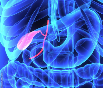
If you have just heard the words “type 4 choledochal cyst,” you are probably trying to picture something you have never seen and cannot feel. This page walks through what a type IV cyst is, where it sits in the body, how doctors tell IVa from IVb, how it is repaired, and what the years afterward usually look like. It is written for patients and parents, in plain English. Please treat it as general information, not medical advice — your own decisions belong to you and your care team.
What a type 4 choledochal cyst is
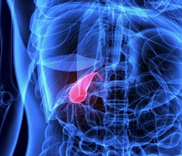
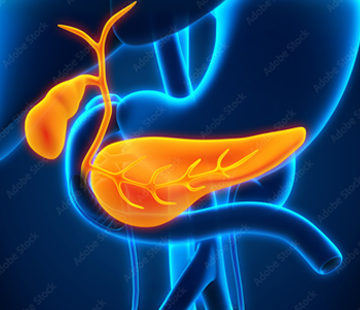
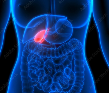
Bile is made in the liver and drains to the intestine through a system of small pipes called the biliary tree. Think of it as a river system. Tiny streams inside the liver (the intrahepatic ducts) join into bigger channels, which leave the liver as the right and left hepatic ducts. Those merge into the common hepatic duct. The gallbladder empties into it through the cystic duct, and from there the pipe is called the common bile duct, which runs down behind the pancreas and empties into the duodenum, the first part of the small intestine.
A choledochal cyst is not a lump growing on the outside of anything. It is a stretched, ballooned section of that pipework itself — the duct wall bulges outward, bile pools instead of flowing briskly, and stones and infection become more likely. The Todani classification sorts these into five types by which section is affected. Type IV is defined by more than one dilated segment, in contrast with type I, which is a single ballooned segment of the common bile duct, or type III, which sits inside the wall of the duodenum. Type IV is generally described as the second most common form.
Type IVa versus type IVb
- Type IVa — multiple dilated segments involving both the ducts inside the liver and the ducts outside it. This is by far the more common of the two and the more demanding to treat, because part of the problem is inside the liver where a surgeon cannot simply cut it out.
- Type IVb — multiple dilated segments confined to the extrahepatic ducts only, outside the liver. The ducts within the liver look normal.
The distinction matters enormously for surgery, so it is worth asking your team, in writing, which subtype is on your imaging. If the dilation is only inside the liver and nowhere outside it, that is not type IV at all — that is type V, or Caroli disease.
How common is it?
Choledochal cysts of all kinds are rare: roughly 1 in 100,000 to 150,000 live births in Western countries, but as frequent as about 1 in 1,000 in parts of Asia. Females are affected about three to four times as often as males. Within that group, type IV accounts for roughly 12% to 35% of all choledochal cysts depending on the series, with type I making up most of the rest. In nearly all patients an abnormal junction between the bile duct and the pancreatic duct is found, which allows pancreatic juice to wash back into the bile duct and is thought to drive the damage.
Typical age and how it shows up
Most choledochal cysts are found in childhood; only about a quarter are discovered in adults, and roughly two-thirds of patients have symptoms before age 10. The textbook triad of pain, jaundice, and a palpable lump in the upper right abdomen appears in only about 20% of cases, so most people do not look like the textbook. Infants and young children more often show jaundice, pale stools, or a mass; older children and adults more often have recurring right upper abdominal pain, sometimes with fever, nausea, or bouts of pancreatitis. Our page on symptoms goes through these in more detail.
Diagnosis and imaging
Ultrasound is almost always first: it is quick, harmless, and detects choledochal cysts with a sensitivity of about 71% to 97%, though it often cannot explain the cause. MRCP — a specialised MRI of the bile ducts, with no radiation — is considered the gold standard, with a reported sensitivity of 90% to 100%, and it is the study that maps the intrahepatic ducts well enough to separate IVa from IVb. CT cholangiography is around 90% sensitive. A HIDA scan is close to 100% sensitive for type I but drops to about 67% for type IVa, because it shows flow rather than detailed anatomy. ERCP gives the highest diagnostic accuracy but carries real risks of pancreatitis and cholangitis, so it is generally reserved for cases where treatment may be done at the same time. See diagnosis for what each test feels like.
Treatment
Type IVa
The core operation is complete excision of the diseased extrahepatic duct — the cyst is removed rather than merely drained — followed by a Roux-en-Y hepaticojejunostomy, in which a limb of small intestine is brought up and sewn to the healthy duct at the liver so bile has a fresh route to the gut. The gallbladder is removed as part of the same operation. The intrahepatic dilation is usually left alone if it is causing no trouble. Partial hepatectomy — removing a segment or lobe of liver — is added when the intrahepatic disease is localised and problematic: stones inside the liver ducts, a tight intrahepatic stricture, an abscess, or a lobe that is already damaged. Simply draining a cyst into the bowel without removing it is no longer acceptable practice; that approach carries roughly a fourfold higher cancer risk than excision.
Type IVb
Because the liver ducts are normal, IVb is treated like type I: full excision of the affected extrahepatic ducts plus Roux-en-Y hepaticojejunostomy, increasingly by laparoscopic or robotic approach. No liver resection is needed. More detail is on our treatments page.
Complications and cancer risk, honestly
Across all choledochal cysts, about 11% of patients develop a biliary malignancy in the largest meta-analysis, and reported cholangiocarcinoma rates in the literature range widely, from around 5% to 28% depending on the series. Risk climbs with age — the median age at cancer diagnosis in cyst patients is around 49.5 years — and is very low in children. When cancer does occur, outcomes are poor, which is precisely why complete removal before cancer develops is the whole point of surgery.
Type IVa specifically carries the highest complication rate of any type after surgery, mainly intrahepatic duct stones and narrowing (stricture) at the join between duct and bowel. Adults have significantly higher rates of postoperative stricture than children. Even so, results are good overall: series report about an 89% event-free rate and five-year survival around 95.5%, and one laparoscopic series of 110 patients with type I and IVa cysts reported a 10% overall complication rate. Our complications page covers cholangitis, stones, pancreatitis, and strictures in plain terms.
Long-term follow-up
- Lifelong surveillance, because a residual risk of cholangiocarcinoma and gallbladder cancer persists even after complete excision — a fact worth knowing without letting it dominate your life.
- Periodic liver blood tests, and imaging (usually ultrasound or MRCP) at intervals your hepatologist sets.
- A low threshold for seeking care with fever, pain, or jaundice, which can signal cholangitis or a stricture.
- For type IVa, particular attention to the intrahepatic ducts left behind.
Our post-operation page describes recovery and what normal looks like afterward.
What this means for you
Type IV is a more involved diagnosis than type I, and IVa in particular means a longer conversation with your surgeon and a longer follow-up horizon. But it is a fixable problem with a well-established operation, most people do well, and the surveillance that follows is routine rather than ominous. Ask which subtype you have, ask whether any liver resection is planned and why, and ask who will follow you in ten years’ time. If it helps to hear from families who have already been through it, visit find community, or use find doctors to locate a specialist centre.
Sources
- Choledochal Cyst — StatPearls, NCBI Bookshelf
- Choledochal Cysts: Background, Pathophysiology, Epidemiology — Medscape
- Choledochal Cysts Treatment & Management — Medscape
- Choledochal Cyst — Radiopaedia
- Long-term complications after extrahepatic cyst excision for type IV-A choledochal cysts — PubMed Central
- Choledochal Cyst: Types, Causes & Treatment — Cleveland Clinic