Choledochal Cysts Type V
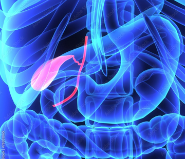
Type 5 choledochal cyst has its own name — Caroli disease — and it behaves differently enough from the other types that it is often discussed as a liver condition rather than a bile duct one. This page explains what Caroli disease is, how it differs from Caroli syndrome, why kidneys and genetics come into the conversation, and what treatment and long-term monitoring involve. It is general information, not medical advice.
What type V is, and where it sits
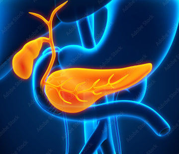
Picture the biliary tree as a river system draining the liver. Small streams inside the liver gather into larger channels; those leave the liver and become the common hepatic duct, then the common bile duct, which carries bile down to the intestine. In type I and most other types, the ballooned section is on the trunk outside the liver. In type V, the dilations are entirely inside the liver: segmental, sac-like widenings of the large intrahepatic bile ducts, often multiple, sometimes clustered in one lobe and sometimes scattered throughout. The ducts outside the liver are normal. That single anatomical fact drives everything else — you cannot simply cut out a duct that runs through liver tissue, so treatment is about liver segments rather than duct reconstruction.
If dilation involves both the ducts inside the liver and those outside it, that is type IVa, not type V. The distinction is made on imaging and is worth confirming, because the operations are different. The full classification is on our types overview.
Caroli disease versus Caroli syndrome
- Caroli disease (sometimes called simple or isolated Caroli disease) — dilated intrahepatic ducts alone, with no scarring of the liver. The dominant problem is bile sitting still: sludge, stones, and repeated episodes of cholangitis (bile duct infection).
- Caroli syndrome — the same duct dilation plus congenital hepatic fibrosis, a condition in which bands of fibrous scar tissue run through the liver from birth. Those bands squeeze the small portal veins and raise pressure in the portal circulation. This is called non-cirrhotic portal hypertension, and it can lead to an enlarged spleen, low platelet counts, fluid in the abdomen, and bleeding from varices in the oesophagus.
Both conditions arise from the same underlying developmental problem, a ductal plate malformation — the bile ducts simply do not remodel correctly as the liver forms before birth. Caroli syndrome is linked to changes in the PKHD1 gene on chromosome 6, the same gene responsible for autosomal recessive polycystic kidney disease (ARPKD). PKHD1 makes a protein called fibrocystin that is needed for normal development of both bile ducts and kidney tubules, which is why liver and kidney disease travel together. Caroli syndrome is inherited in an autosomal recessive pattern, meaning both parents carry a change without being affected. Isolated Caroli disease is usually sporadic — not inherited from either parent — though it has occasionally been reported in families and in association with autosomal dominant polycystic kidney disease.
How common is it?
Type V is consistently the rarest presentation to encounter in person, even though older classification tables have quoted it as up to 20% of all choledochal cysts. In absolute terms, isolated Caroli disease is estimated at about 1 in 1,000,000 people. Figures for Caroli syndrome vary by source: it is put at roughly 1 in 100,000 by the National Organization for Rare Disorders, while StatPearls cites 1 in 10,000 to 20,000 live births. Congenital hepatic fibrosis on its own is estimated near 1 in 20,000 live births. Caroli syndrome is generally the more frequently encountered of the two. Unlike other choledochal cysts, which affect females three to four times as often, Caroli disease affects males and females about equally, and it is reported more often in Asian populations.
Typical age and presentation
Over 80% of patients have symptoms and are diagnosed before the age of 30, though a first episode can come at any age and some people are diagnosed incidentally on a scan done for something else. Simple Caroli disease often declares itself in adolescence or young adulthood with the classic cholangitis picture: fever, right upper abdominal pain, and jaundice, sometimes with itching. Others have chronic, grumbling right-sided pain without dramatic attacks. Caroli syndrome may show up much earlier — in infancy alongside ARPKD — or in later childhood and adolescence with signs of portal hypertension such as a large spleen, low blood counts, or a variceal bleed. Our symptoms page describes what these episodes actually feel like.
Diagnosis and imaging
Blood tests are often unremarkable between attacks; alkaline phosphatase and bilirubin rise during obstruction or infection, and low white cells or platelets can hint at portal hypertension. Imaging makes the diagnosis:
- Ultrasound — cheap and safe, shows saccular dilations inside the liver and is usually the first test.
- CT — maps focal or diffuse dilation and can show fibrosis and varices.
- MRCP — the most commonly used and generally preferred study, because it shows the whole biliary tree and how much of the liver is involved without radiation, and is safer than ERCP, which carries a risk of pancreatitis and infection.
- The central dot sign — on contrast CT or MRI, a small bright dot sitting inside a dilated duct. That dot is the portal vein branch and its fibrous sheath, surrounded by the ballooned duct. It is highly suggestive of Caroli disease, though not completely exclusive to it.
Kidney imaging and, where appropriate, genetic testing are usually part of the workup too. See diagnosis for more.
Treatment
Care is staged. Acute cholangitis is treated with antibiotics covering gram-negative and anaerobic bacteria, with drainage by ERCP stent or a percutaneous drain when a duct is blocked. Ursodeoxycholic acid is used to help bile flow and reduce stone formation. Portal hypertension in Caroli syndrome is managed on its own terms, with variceal screening and treatment.
Surgery is guided by how much liver is involved:
- Unilobar (one-lobe) disease — segmental resection or lobectomy removes the affected liver and, with it, the source of infection and stones. The left lobe is the one most often involved. Long-term outcomes are excellent, with reported five-year survival around 97% after partial hepatectomy, and many people have no further episodes.
- Diffuse or bilobar disease — resection is not feasible and outcomes are poor if attempted. Liver transplantation is the only curative option and is considered for recurrent cholangitis, biliary cirrhosis, liver failure, or severe portal hypertension. Reported five-year survival after transplant is around 89%. Combined liver-kidney transplantation is sometimes needed when ARPKD has damaged the kidneys.
Our treatments and post-operation pages cover what to expect around surgery.
Complications and cancer risk
The everyday complications are recurrent cholangitis, intrahepatic stones, and occasionally liver abscess or sepsis; in Caroli syndrome, add portal hypertension and its consequences. The most serious long-term concern is cholangiocarcinoma, bile duct cancer, with reported prevalence in Caroli disease and syndrome of about 7%, and up to 15% in some series — far above the general population, and driven by decades of bile stasis and inflammation. More on complications here.
Long-term follow-up
European guidance suggests MRCP every 12 months after diagnosis to screen for cholangiocarcinoma, though this is a weak recommendation reflecting limited evidence. Blood tumour markers such as CA 19-9 and CEA perform poorly and are not recommended for routine monitoring on their own. Beyond that, follow-up usually includes liver function tests, variceal screening if there is portal hypertension, kidney monitoring where ARPKD is present, and a plan for prompt antibiotics at the first sign of cholangitis. Care is best delivered by a team — hepatologist, transplant surgeon, radiologist, and, for children, a paediatric specialist. Our find a doctor page can help you locate centres with experience, and current research tracks what is changing.
What this means for you
A Caroli diagnosis is heavy news, largely because it is lifelong rather than a one-off repair. But the outlook is genuinely better than the rarity of the name suggests: people with disease confined to one lobe are often cured by resection, and those with more extensive disease can live full lives with good infection control, regular monitoring, and transplantation held in reserve. The two questions worth asking early are how much of your liver is involved, and whether congenital hepatic fibrosis is present — those answers shape everything that follows. You are not the only family navigating this; find community to connect with others.
Sources
- Caroli Disease — StatPearls, NCBI Bookshelf
- Caroli Disease — National Organization for Rare Disorders
- Congenital Hepatic Fibrosis, Caroli Disease and Caroli Syndrome — AASLD Liver Fellow Network
- Caroli Disease: Symptoms, Diagnosis, Treatment and Outlook — Cleveland Clinic
- Choledochal Cyst — StatPearls, NCBI Bookshelf
- Choledochal Cysts Treatment & Management — Medscape